


Researchers in the US have designed a wearable ultrasound monitor that can image organs within the body without the need for an ultrasound operator or application of gel.
The patch could help patients with bladder or kidney disorders more easily track whether these organs are functioning properly, the researchers said.
The approach could also be adapted to monitor other organs within the body by changing the location of the ultrasound array and tuning the frequency of the signal.
Such technology could potentially enable earlier detection of cancers that form deep within the body, such as ovarian cancer.
Study senior author Canan Dagdeviren, an associate professor in MIT’s Media Lab, said: “Millions of people are suffering from bladder dysfunction and related diseases, and not surprisingly, bladder volume monitoring is an effective way to assess your kidney health and wellness.”
“This technology is versatile and can be used not only on the bladder but any deep tissue of the body.
“It’s a novel platform that can do identification and characterisation of many of the diseases that we carry in our body.”
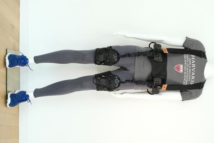
The researchers created a flexible patch made of silicone rubber, embedded with five ultrasound arrays made from a new material that the researchers developed for this device.
The arrays are positioned in the shape of a cross, which allows the patch to image the entire bladder, which is about 12 by 8 centimetres when full.
The material that makes up the patch is naturally sticky and adheres gently to the skin, making it easy to attach and detach.
Once placed on the skin, underwear or leggings can help to hold the patch in place.
For the study, the researchers recruited 20 patients with a range of BMIs.
Participants were first imaged with a full bladder, then with a partially empty bladder, and then with a completely empty bladder.
The images obtained from the new patch were similar in quality to those taken with traditional ultrasound, and the ultrasound arrays worked on all subjects regardless of their BMI.
With this patch, no ultrasound gel is needed, and no pressure needs to be applied, as with a regular ultrasound probe, because the field of view is large enough to encompass the entire bladder.
To see the images, the researchers connected their ultrasound arrays to the same kind of ultrasound machine used in medical imaging centres.
However, the MIT researchers are now working on a portable device, about the size of a smartphone, that could be used to view the images.
Anthony E. Samir is director of the MGH Center for Ultrasound Research and Translation and Associate Chair of Imaging Sciences at MGH Radiology.
The researcher said: “In this work, we have further developed a path toward clinical translation of conformable ultrasonic biosensors that yield valuable information about vital physiologic parameters.
“Our group hopes to build on this and develop a suite of devices that will ultimately bridge the information gap between clinicians and patients.”
Image: MIT