
By Angel Alberich-Bayarri, CEO of Quibim
For decades, the biopsy has been the gold standard of cancer diagnosis, an essential step that gives clinicians the microscopic truth about a tumour.
It is precise, but by its nature also invasive, limited to certain moments in a patient’s journey, and not always feasible depending on tumour location or patient condition.
Now, in the era of artificial intelligence (AI), we are entering a new chapter: one where the only question is not “biopsy or not,” but how to end the uncertainty in the many situations where a biopsy alone cannot give the full picture.
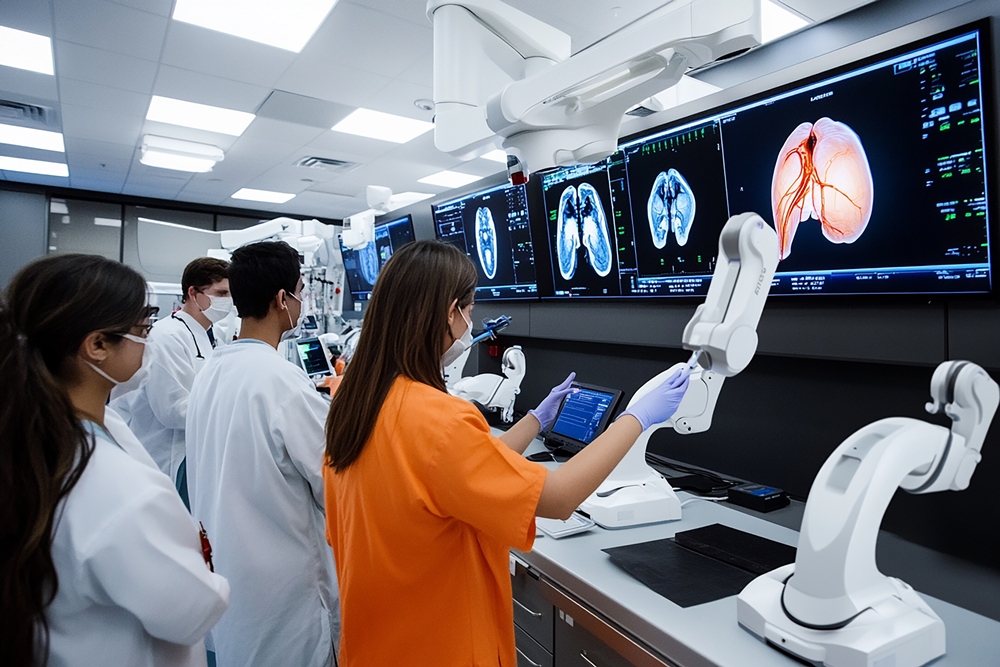
AI-powered imaging applied to MRI, CT, PET images is becoming a powerful ally to pathology, able to detect and characterise tumours with a level of biological insight once thought possible only from tissue samples.
By analysing medical images with advanced deep learning models, we can turn pixels into biomarkers, revealing the biology of disease without an additional incision or needle.
Recent research, for example, shows that combining advanced image analysis with AI can help predict key cancer marketers – such as PD-L1 status in lung cancer – directly from CT scans.
PD-L1 is an important indicator for deciding whether a patient is likely to benefit from immunotherapy, and today it can only be measured through a tissue biopsy.
While still in the research stage, these AI-based approaches could soon help clinicians assess biomarker status more rapidly, safely, and at scale.
From single events to continuous insight
Biopsies usually happen once, at diagnosis or a key decision point, but cancer evolves over time. Tumour biology can change between diagnosis, treatment, and relapse.
AI-enabled imaging complements the biopsy by filling those gaps:
- In patient follow-up, where repeated biopsies would be too risky or impractical
- In population screening, where sampling everyone is impossible
- In hard-to-reach tumours, where biopsy may not be feasible at all
The result is a more complete, longitudinal view of the disease, and less time spent in uncertainty for both patients and clinicians.
Beyond individual cases: expanding access and accelerating trials
In many parts of the world, the infrastructure for timely, high-quality biopsies is scarce. Imaging, however, is often more widely available.
With AI, those scans become more than pictures, they become sources of actionable biological data, helping clinicians make better decisions even where pathology services are limited.
The same applies to research.
Non-invasive imaging biomarkers can make clinical trials more inclusive by allowing participation of patients who cannot undergo a biopsy due to tumour location, comorbidities, or other constraints.
This accelerates evidence generation, reduces trial delays, and promotes equity by including patients who would otherwise be excluded due to biopsy restraints.
Earning trust and ensuring impact
The power of imaging AI comes with responsibilities: rigorous validation across diverse populations and imaging systems, transparent methods, and close collaboration among clinicians, data scientists, and regulators.
Trust must be built step-by-step, just as it was for every major medical innovation in the past century.
A new diagnostic paradigm
This is not about replacing biopsies, it’s about completing the picture.
By combining the strengths of pathology and imaging AI, we can reduce the uncertainty that delays decisions, causes anxiety, and limits treatment options.
The future of cancer diagnosis is one where patients benefit from both the microscopic truth of the biopsy and the continuous, non-invasive insights of imaging, a future where clarity comes faster, safer, and for more people.